3401 Civic Center Blvd
Philadelphia, PA, 19104
(215) 590-4545
Medical Illustration
Your Custom Text Here

Your Custom Text Here
The complexity of the body' structure and its function is best described when strong clear visuals are accompany written text. The following examples focuses on such examples.
The blood supply to the neck of the femur has been poorly understood for years. This illustration tracks the the course of the arteries and highlight their relationship to the muscles of the hip.
The complexity of the human knee is only matched by its beauty.
The two illustrations shown here provide an anterior (frontal) view and an axial (top) view of the knee joint. Particular attention is given to the ligaments and tendons of the joint.
This is illustration shows the section of the knee joint that sits atop the tibia bone.
In July 2015 The Children's Hospital of Philadelphia performed the first ever bilateral hand transplant on a child. I was fortunate enough to illustrate the procedure. The illustrations were used to educate the public about the steps of the procedure and were used as part of the hospital's press release.
A pre surgery view of the patient and his arms.
This is Zion's story. It chronicles his journey of getting two new hands. My illustrations are used to bring clarity to the surgical procedure.
Bone plates are used to mechanically attach the bones of the forearm. Care was taken to preserve the growth plate in the wrist, so the arm will continue to grow with the patient.
Returning blood flow to the donor hands is essential. Attaching the major arteries keeps the tissue alive.
After the veins and nerves are reattached, over a dozen muscles and tendons are connected.
The final step in the procedure is to close the skin.
After a surgery that lasted more than half a day, the patient has new hands.
As technology advances, so too does the ability to develop new and cutting edge surgical techniques which aid in the speed of recovery, aid in safety, and provide outcomes that were before unatainable.
When conjoined twins are joined at the head they are referred to as caraniopagus. This series of illustrations was created to help describe the specifics of the the twins anatomy and how they were successfully separated.
This illustration was incorporated into the press release as part of the artificial womb research at The Children's Hospital of Philadelphia. The illustration posted on a vast array of news organizations around the world. This image was pulled from home page of National Public Radio (NPR).
When a fetus is diagnosed with a myelomeningocele (MMC), or more commonly known as Spina Bifida there is now a procedure which can give some families a chance at improved outcomes and a potentially higher quality of life for the child. This procedure occurs when the fetus is still developing in the the womb and requires the highest level of surgical technique. This series of illustrations created for a fetal surgical atlas, and is primarily drawn in graphite.
This axial cross section shows the defect prior to surgery, the creation of the myofacial flap, and the final sutured closure.
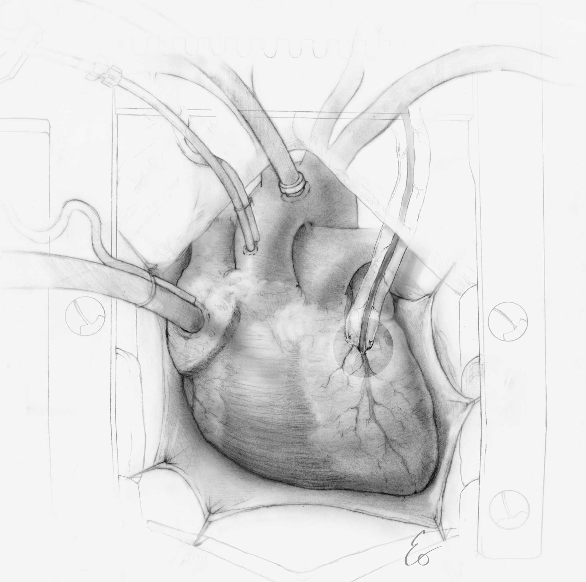
When the coronary arteries of the heart become clogged, it then may be time for a bypass procedure. This graphite illustration shows the surgical view of the procedure including the grafted artery to the heart, and the cannulation, which carries blood to and from the oxigenator.
The VEPTR device is used to treat thoracic insufficiency syndrome (TIS). TIS prevents normal development and function of the lungs. The VEPTR device is used to separate and expand the ribs of the ribcage to create volume and in the process the device can additionally help straighten the spine in cases of extreme scoliosis.
This composition describes the concept of how gene editing is studied to further advance the treatment of genetic disorders while the patient is in the womb.
The cover illustration shows a reimagined concept of how blood in the fetal heart "sculpts" the aortic valve.
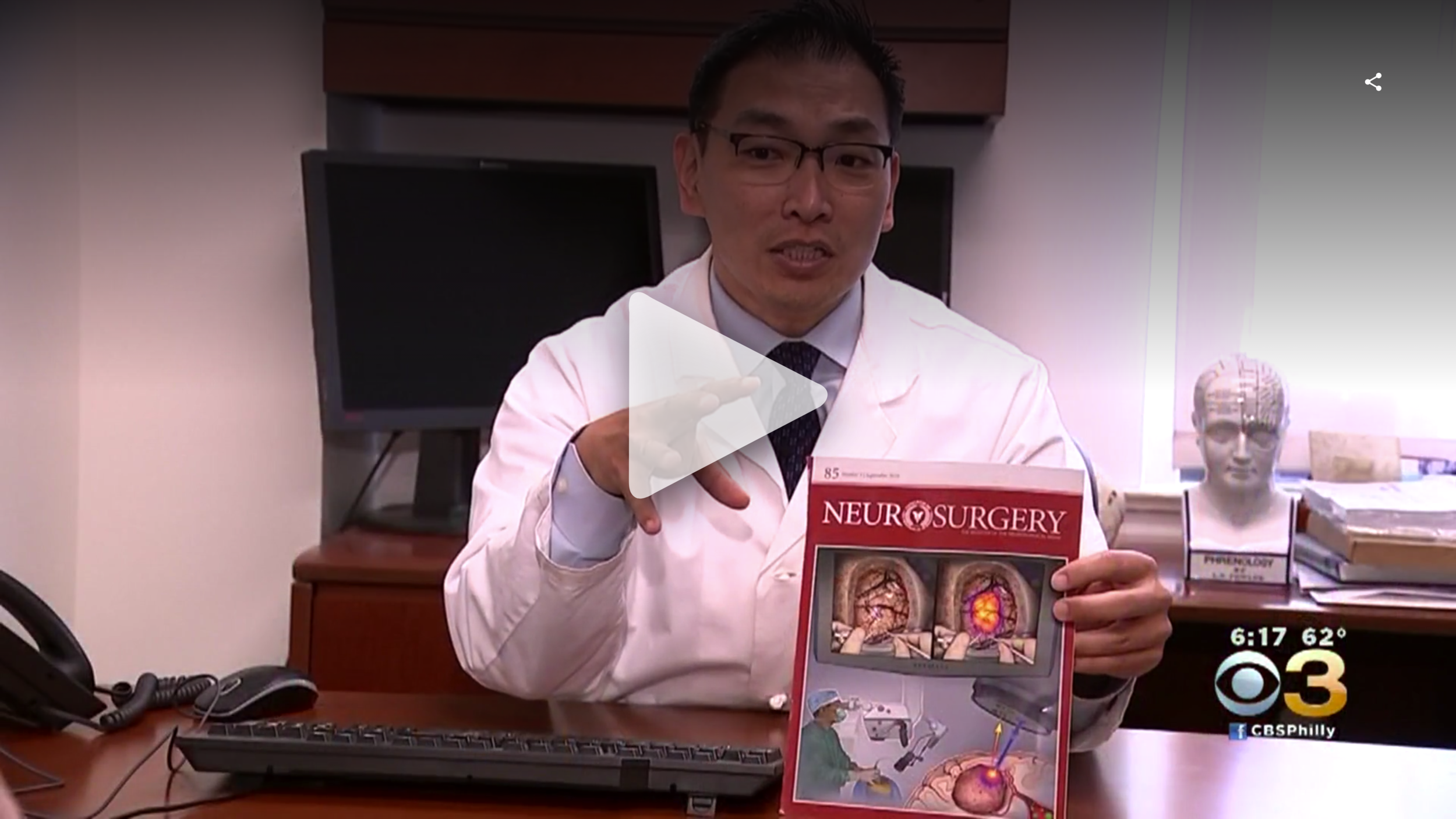
The cover composition for the May 2017 issue of the Journal of Neurosurgery (JNS) shows the key elements of a novel endoscopic approach for treating trigeminal neuralgia. The illustration was created in collaboration with Dr. John Lee at the Hospital of the University of Pennsylvania.
This news piece highlights my work and the brilliant work of Dr. John Lee at Penn medicine.
Stylus is the publication at the University of Pennsylvania which covers the crossroads of medicine, art, and literature. This illustration was awarded the cover in 2015.
This illustration describes the anatomy of the spine and surrounding tissue of patients with this condition.
The treatment for hydrocephalus is to implant a shunt into the expanded ventricle and drain the fluid into the abdomen.
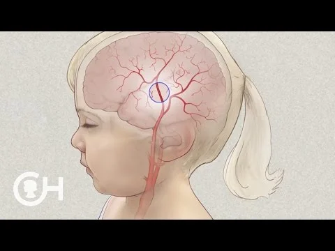
This video from The Children's Hospital of Philadelphia details the condition known as Moya Moya. The series of illustrations seen at minute 3:27 describes the anatomy and surgical procedure.
A Hemispherectomy is the surgical procedure in which one side of the brain is mostly removed. This procedure is used to treat the most extreme types of epilepsy.
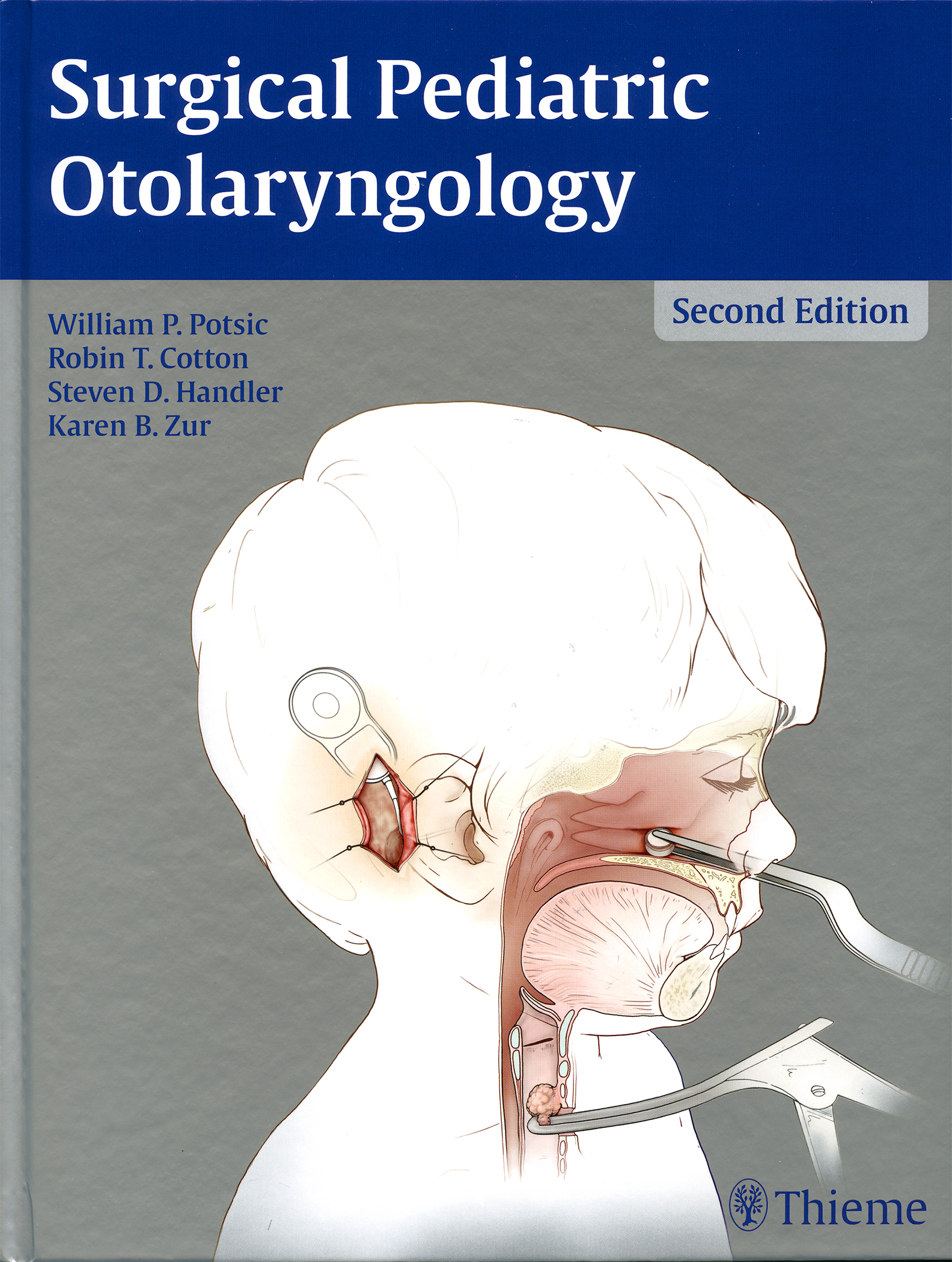
Otolaryngology is the official name for the study of the ear, nose, and throat and is a field of specialization for Eo Trueblood and his studio Stream Studios.
A common treatment is the closed nasal fracture reduction, which requires a long narrow instrument called a Boies Nasal Fracture Elevator to be inserted in the nostril to reposition the displaced bone fragment.
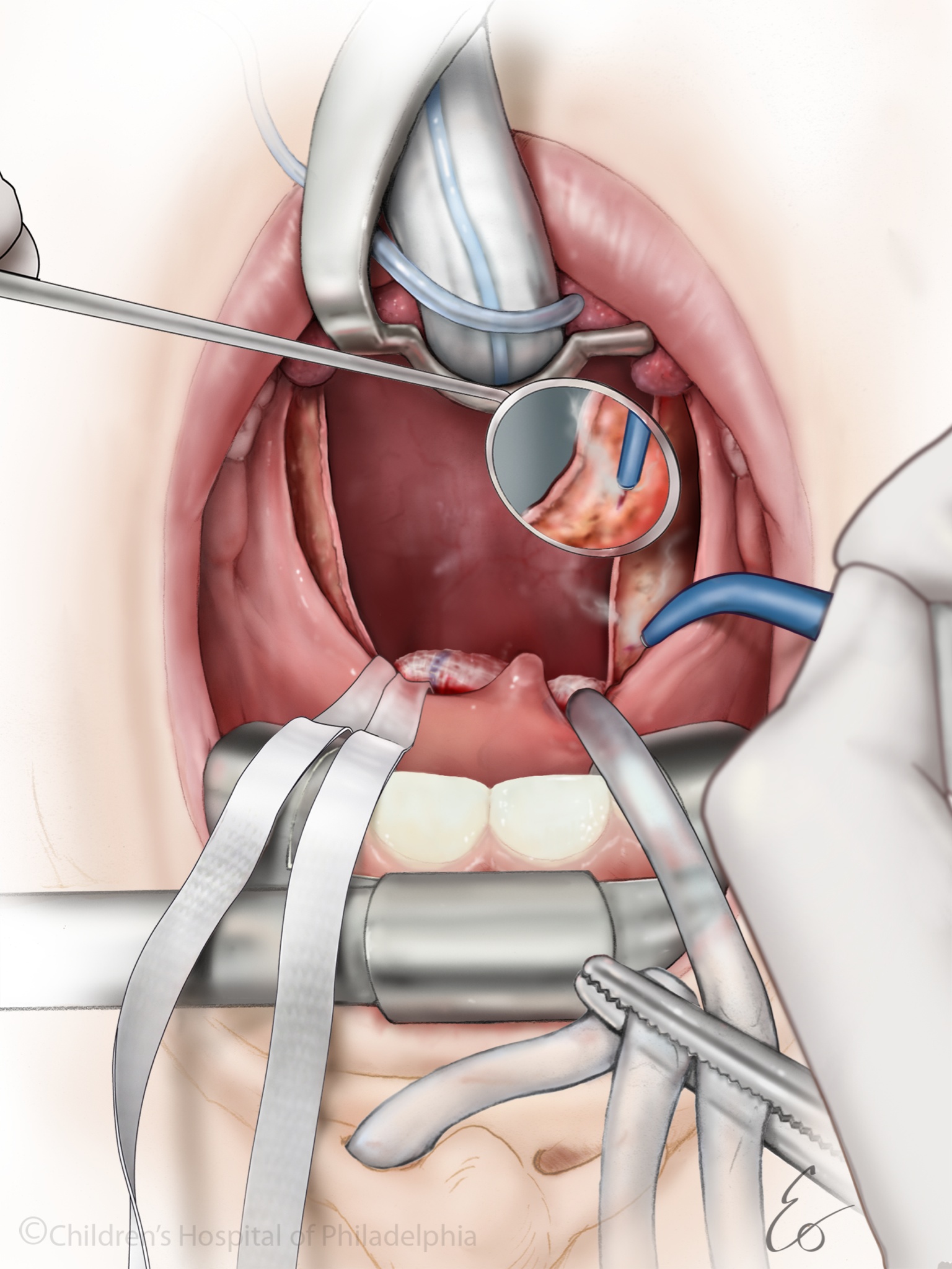
The andenotonsillectomy procedure is comprised of the removal of the tonsils and adenoids. This illustration shows the surgeon using a mirror to inspect the fossa after the removal of the tonsil tissue. The purpose is to cauterize any bleeding vessels to promote healing.
This step in the andenotonsillectomy procedure is to test the size of the adenoid pad in the back of the sinus.
This illustration shows the perspective of the middle ear during Endoscopic Ear Surgery (EES).
The utilization of endoscopes is the next step in the practice of minimally invasive surgeries. Endoscopes are small cameras on the end of long narrow tubes which can be inserted into the body to assist in the visualization of the surgery. Endoscopic surgeries require smaller incisions and often allow for faster recovery times. The gallery shows the endoscope being applied in brain and ear surgery.
Trigeminal neuralgia is a painful and often debilitating condition affecting the facial nerve. The endoscopic approach requires a smaller incision and less manipulation of the brain.
Endoscopic ear surgery is a new technique for visualizing and operating on the the middle ear.
Removal of bone to enlarge opening into the middle ear.
larged external auditory canal, with exposed ossicles of middle ear.
In this case an endoscope provides the surgeon with a view of the surgical area that they would otherwise be unable to see deep inside the patients head.
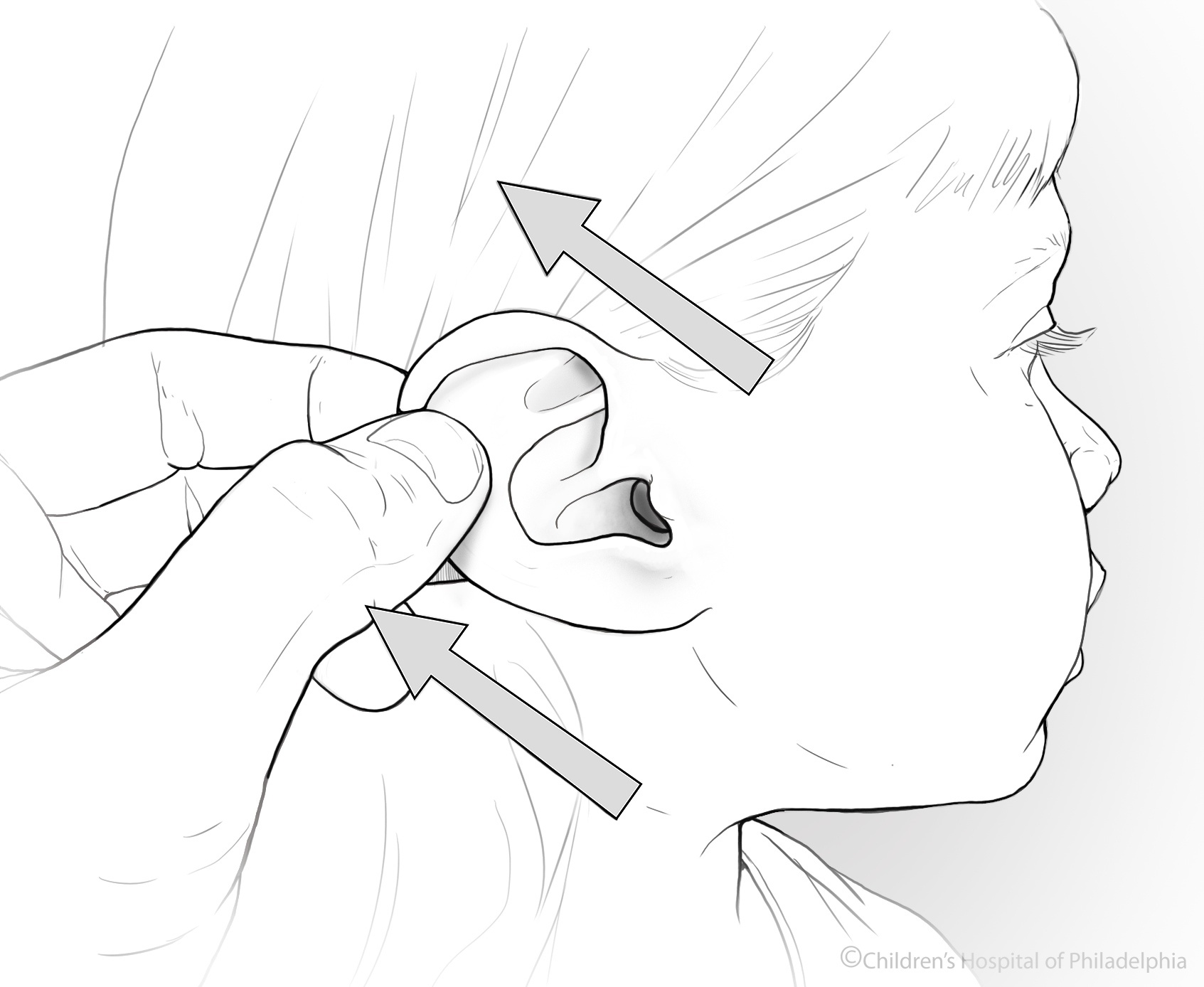
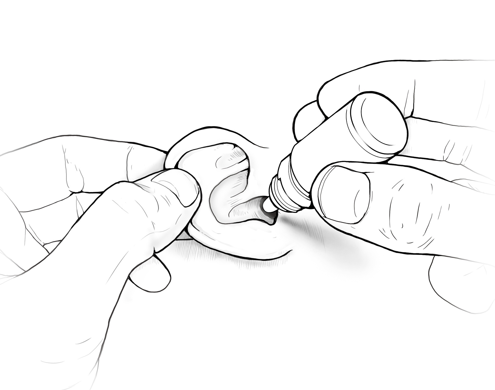
Once a child comes home from the hospital the family becomes the primary care givers tasked with administering medications, maintaining medical devices, and monitoring the health of the child. Education is required to get parents and patients up to speed in terms of knowledge and techniques.
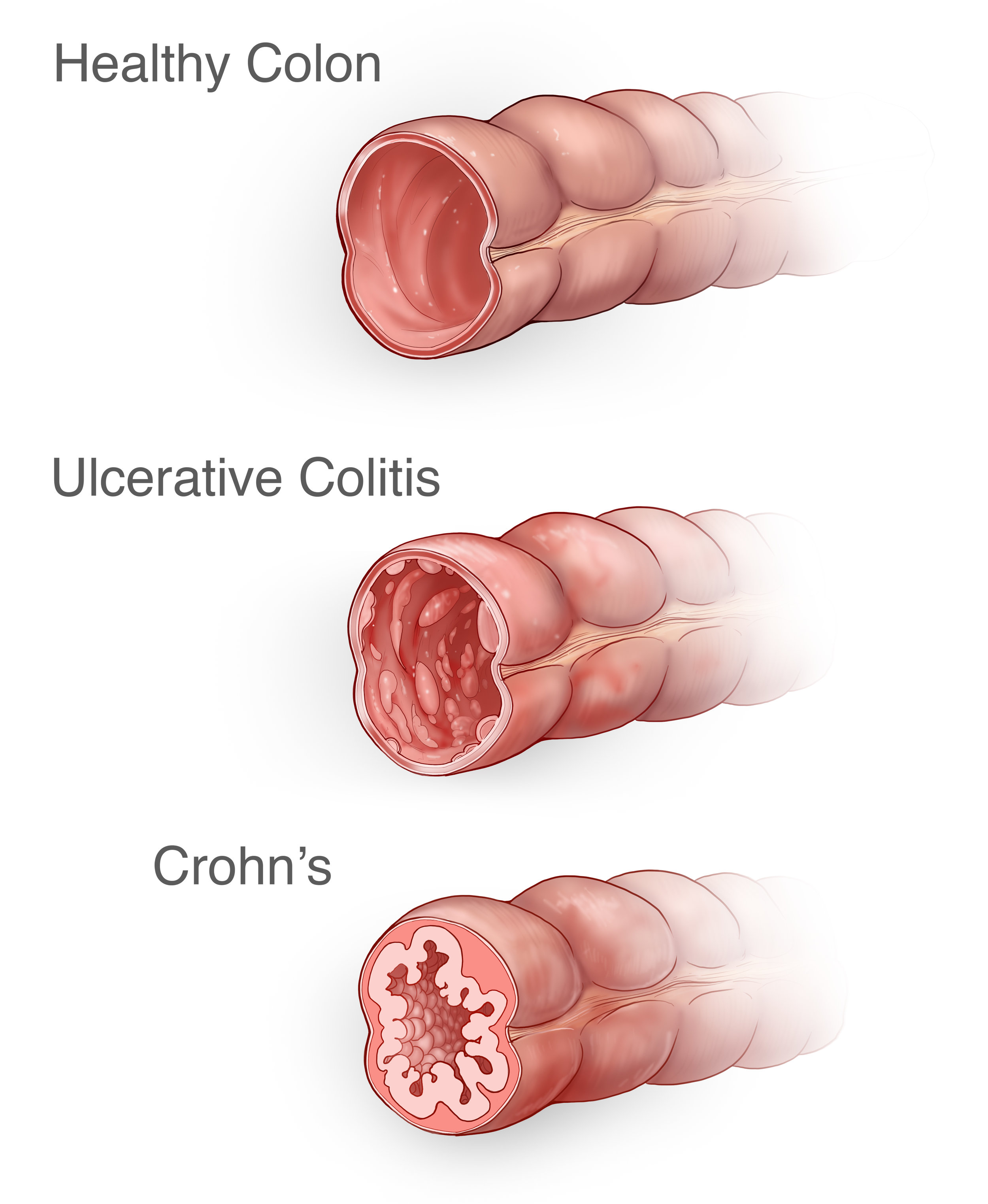
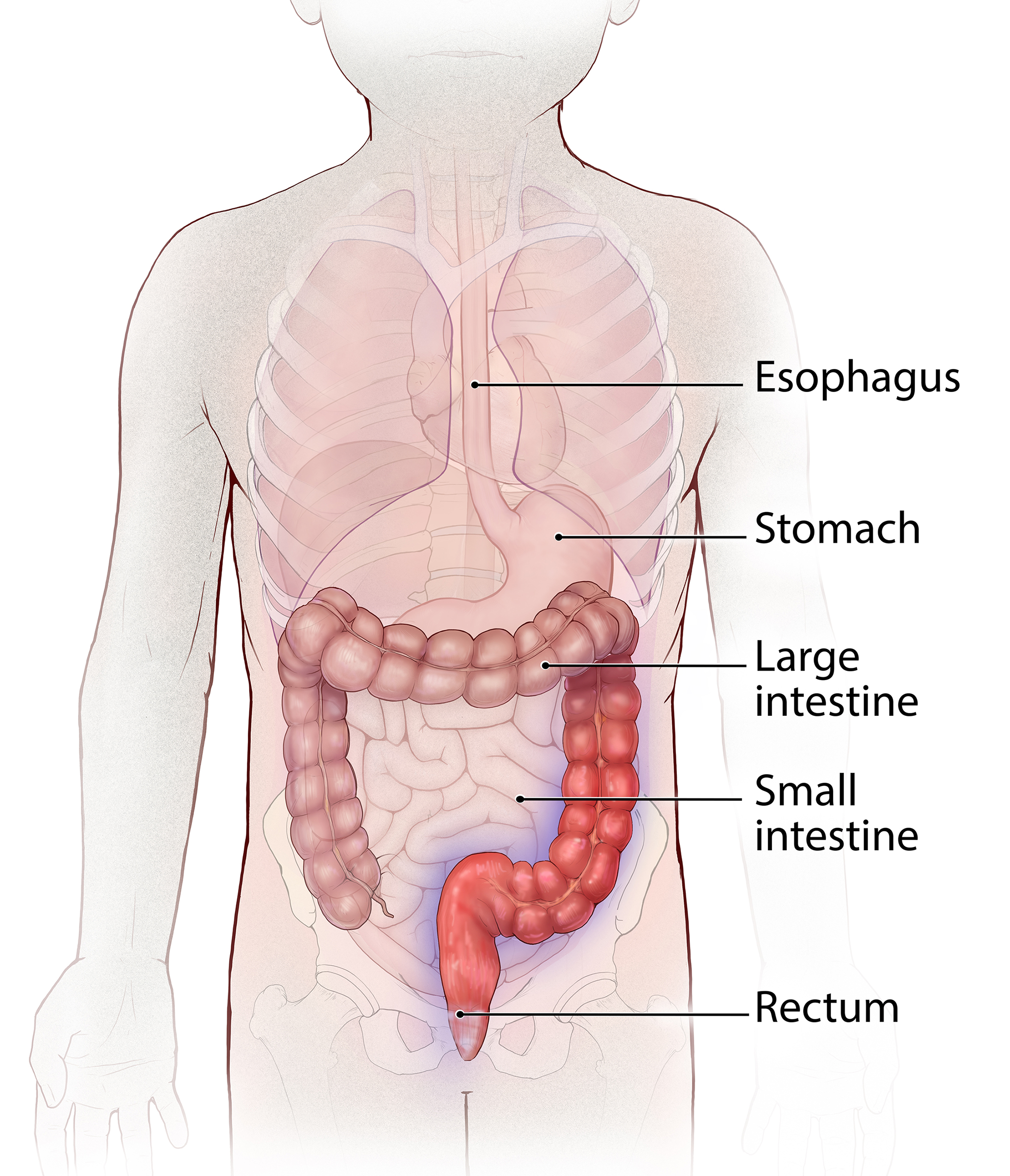
Inflammatory Bowel Disease or IBD is a general term for chronic conditions of the intestines stemming from inflammation and ulcerations. The two most common IBD conditions are Crohn's and Uclerative Colitis.
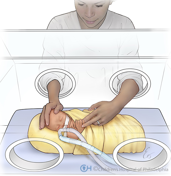
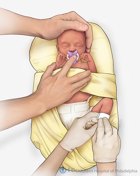
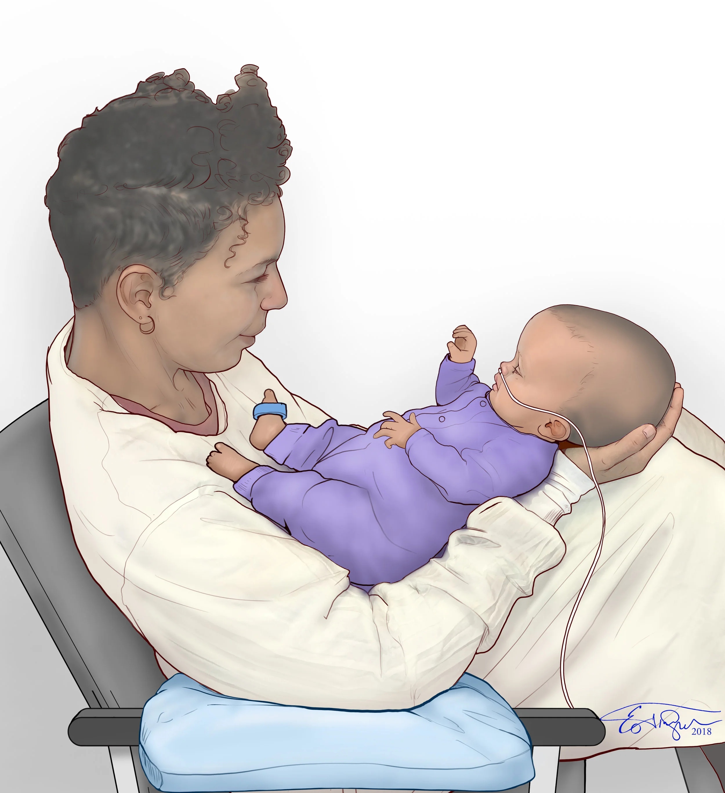
The role of a parent with respects to the care of a preemie, is of the utmost importance. This series of illustrations is part of an instructional program created to aid parents in nurturing their child while in the NICU.
Skin to skin contact (kangaroo care) is key for the health and development of a preemie infant.
Even when an infant is too sick to be picked up, positive touch from a parent or sibling has a beneficial impact.
At times when the infant is being treated by the nurse or doctor, positive touch from the parents, can sooth the infant and reduce distress.
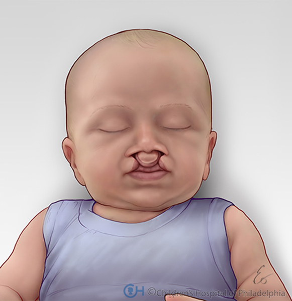
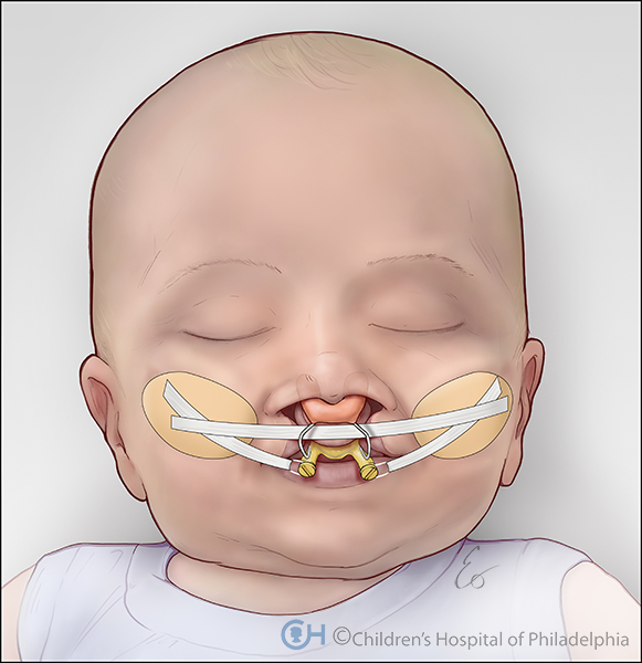
These illustrations were created as part of an educational video on cleft lips and cleft palates for The Children's Hospital of Philadelphia. The third illustration shows the Nasoalveolar Molding (NAM) device as it appears on the patient. The NAM device works to close the cleft which allows for better surgical outcomes.
This illustration shows the unique characteristics of unilateral complete cleft lip and palate.
Unlike the unilateral cleft, the bilateral cleft affects both sides of the child's palate and lip.
The NAM device works to reduce the size of the cleft, increase the surgical outcomes, as well as reduce the number of surgeries over the course of the child's life.